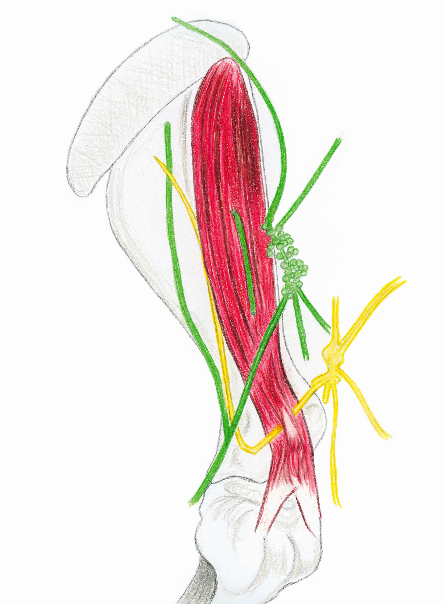
– with the sprascapular nerve (in yellow): it originates from the cranial part of the brachial plexus (the yellow node), reaches the cranial edge of the neck of the scapula, whose lateral face it bypasses to arrive at the deep face of the infraspinatus muscle, going up along the scapular spine. On its way, it provides fibers to the supraspinatus muscle and to the capsule of the scapulohumeral joint. It is the motor of the two above-mentioned muscles and also proprioceptive. Its relationship to the neck of the scapula exposes it to tugging in the event of violent abduction or crushing during strong and prolonged pressure on the shoulder region (for example when restraining a horse on the ground). Its paralysis is translated in the short term by a shoulder which seems to be detached from the thorax and later by the atrophy of the 2 muscles which gives the shoulder a thinned aspect, the scapular spine becoming prominent under the skin.
– with the superficial cervical nodes (in green): often called “prescapular” because of their location at the cranial edge of the shoulder, where they are palpable on the living person (useful when draining the shoulder during massage). Voluminous, this group is made up of 60 to 130 lymph nodes, the largest of which can reach 3 to 4 cm. It is 15 to 30 cm long and 2 to 3 cm wide. Some of its afferent vessels drain the muscles of the superficial and middle planes of the dorsal region of the neck, the lateral aspect of the shoulder, the pectoral region and the forearm, as well as the bones of the shoulder and the thoracic limb.
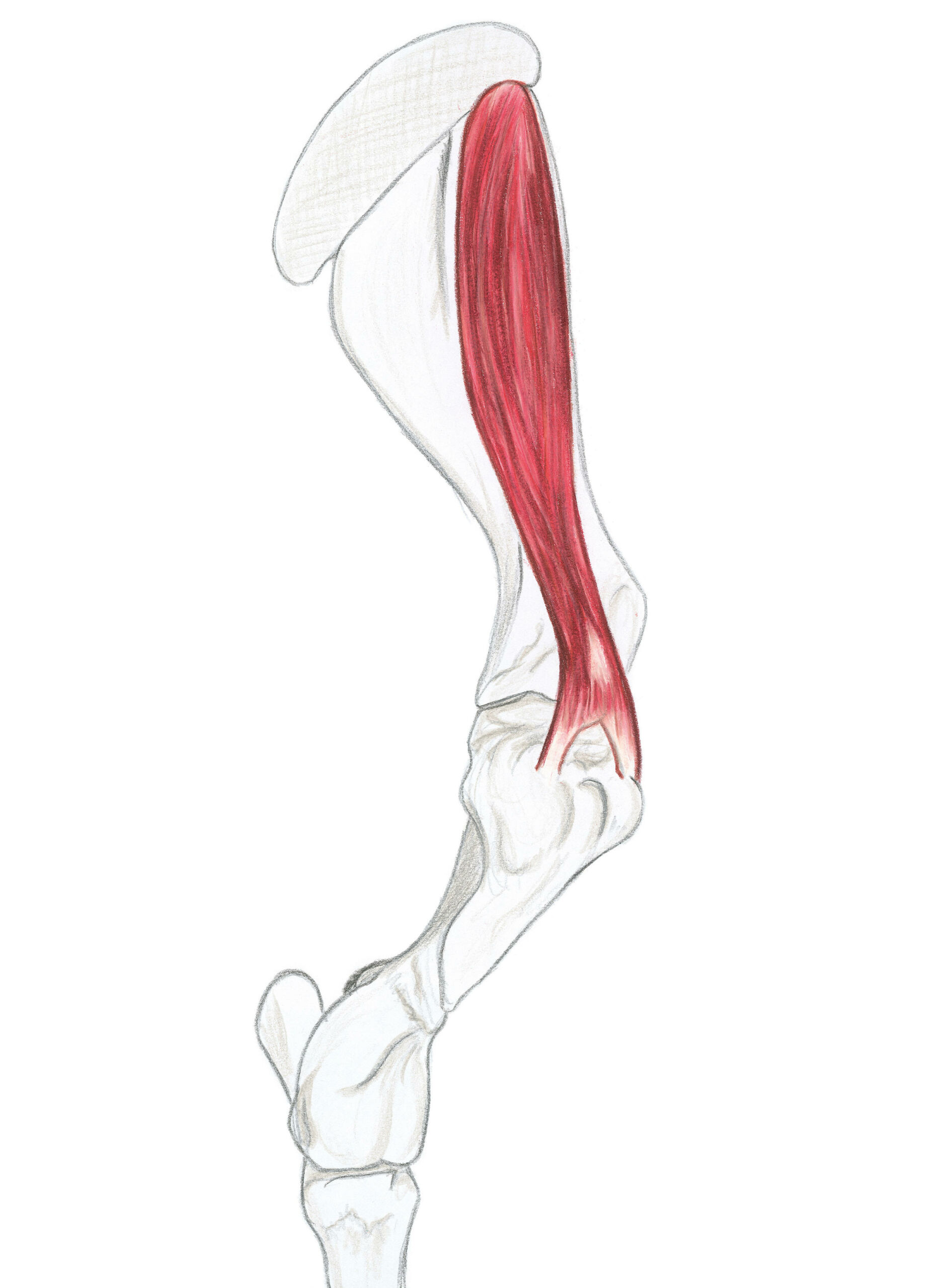
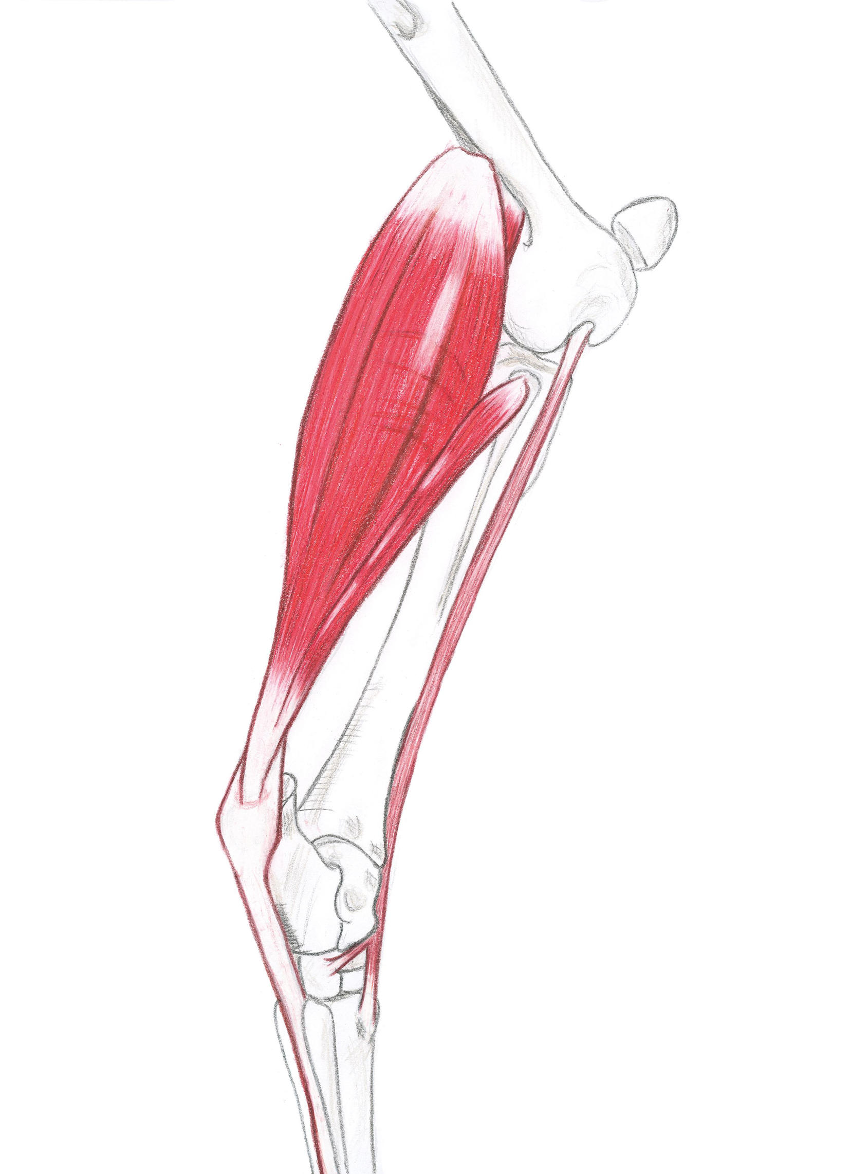
The supraspinatus muscle
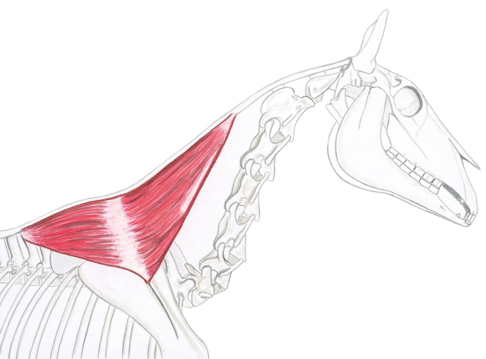
The trapezius muscle
The trapezius muscle
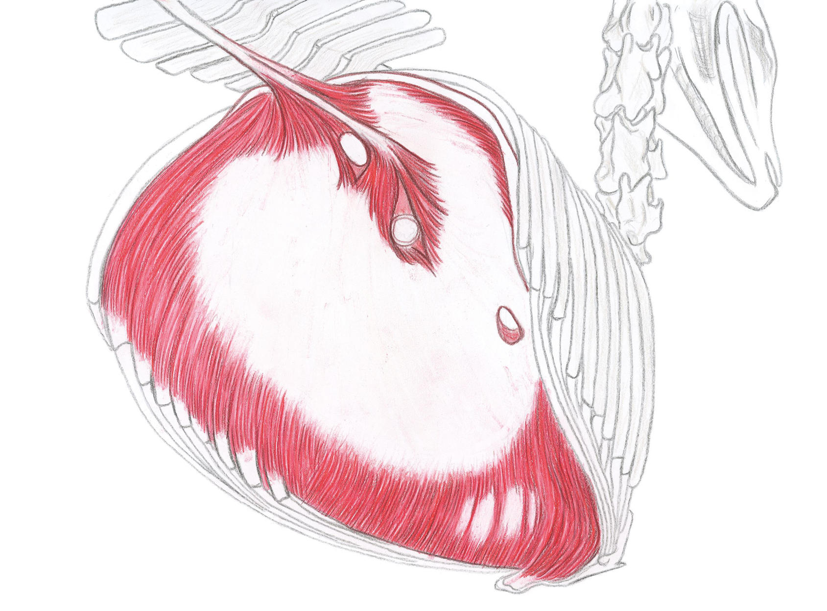
The trapezius muscle: It is a flat, triangular muscle, separated by a strong aponeurosis, located at the level of the scapular spine. It is in two fleshy parts: a cervical part and a thoracic part.
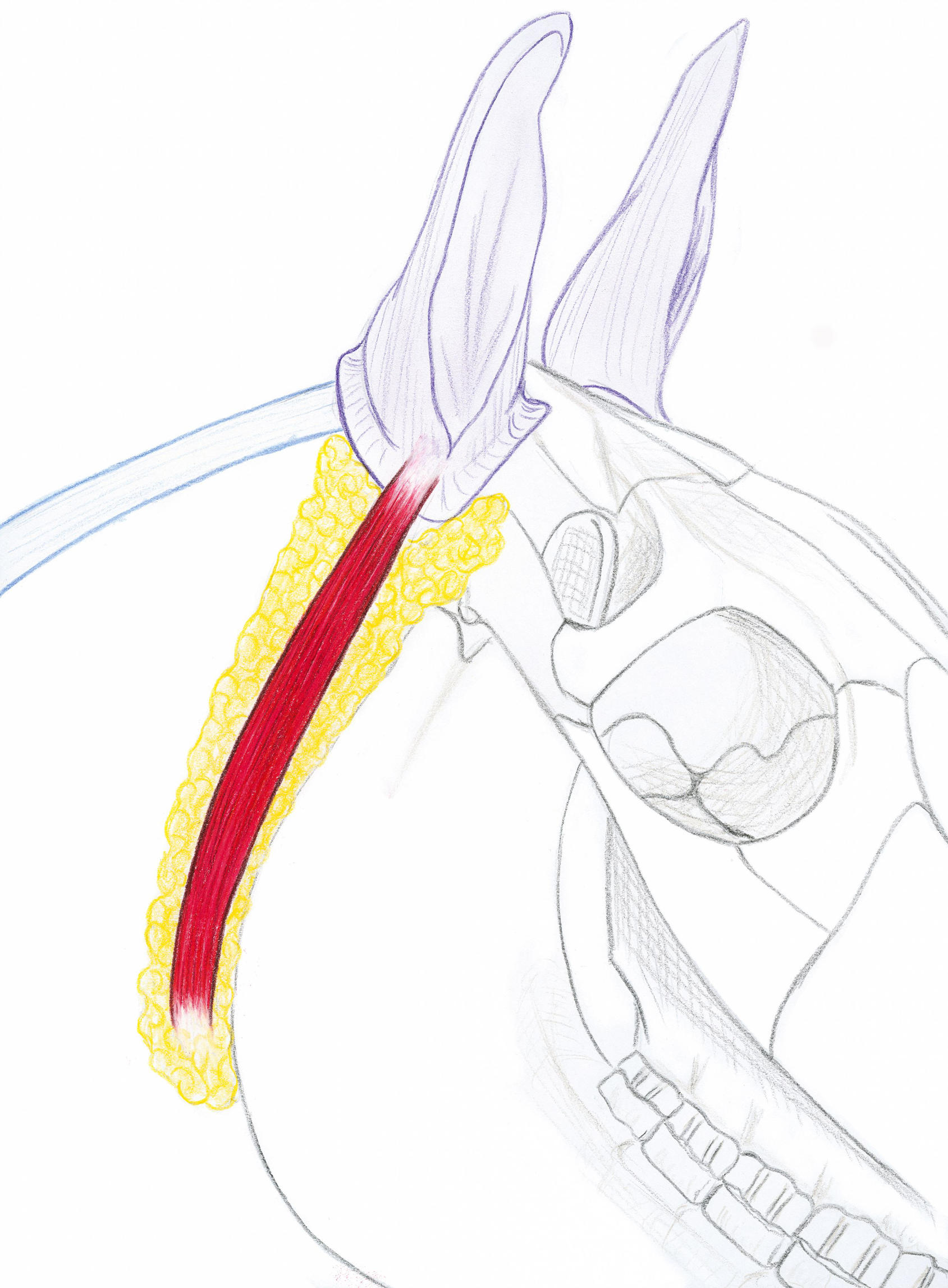
The parotido-auricular muscle
On the surface of the parotid gland, its superficial side is separated from the skin by a thin expansion of the platysma. The deep side covers the parotid gland.
The parotido-auricular muscle
The sterno-hyoid and sterno-thyroid muscles
The sternohyoid and sternothyroid muscles, long and flattened, run along the ventral side of the trachea from the sternal manubrium to the laryngeal region. They are initially joined together and separate at their termination into two parts:
Sternohyoid: ventral side of the body of the hyoid bone.
Sterno-thyroid: caudal edge of the thyroid cartilage blade of the larynx.
The sterno-hyoid and sterno-thyroid muscles
The diaphragm muscle
The diaphragm muscle
The diaphragm is a vast musculoaponeurotic partition that completely separates the thoracic cavity
from that of the abdomen. This complex membrane, thin but solid, has the shape of a very strongly convex dome on the thoracic side. It is maintained tense in this situation by the pressure of the abdominal viscera and by a sort of suction cup action exerted on its cranial face
by the pleural depression. In the vast concavity of the diaphragm are housed, in whole or in part, important viscera: liver, stomach, spleen and in part pancreas and kidneys. The diaphragm has three openings for the aorta, the caudal vena cava and the esophagus.
The interosseous III muscle
The interosseous III muscle
Fibrous in ungulates, where it has long been referred to as the “fetlock suspensory ligament”. It is very strong and entirely formed of white fibrous tissue, within which striated muscle fibers persist. It covers the palmar surface of the main metacarpal/metatarsal bone.
The rectus abdominis muscle of the head
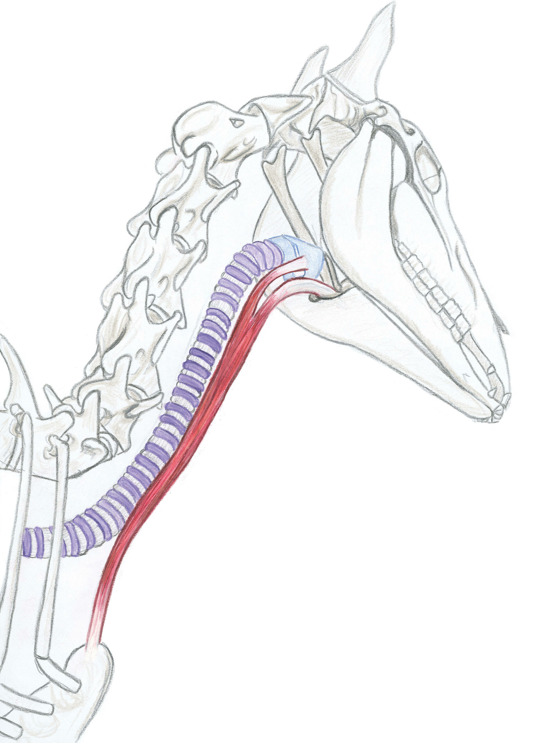
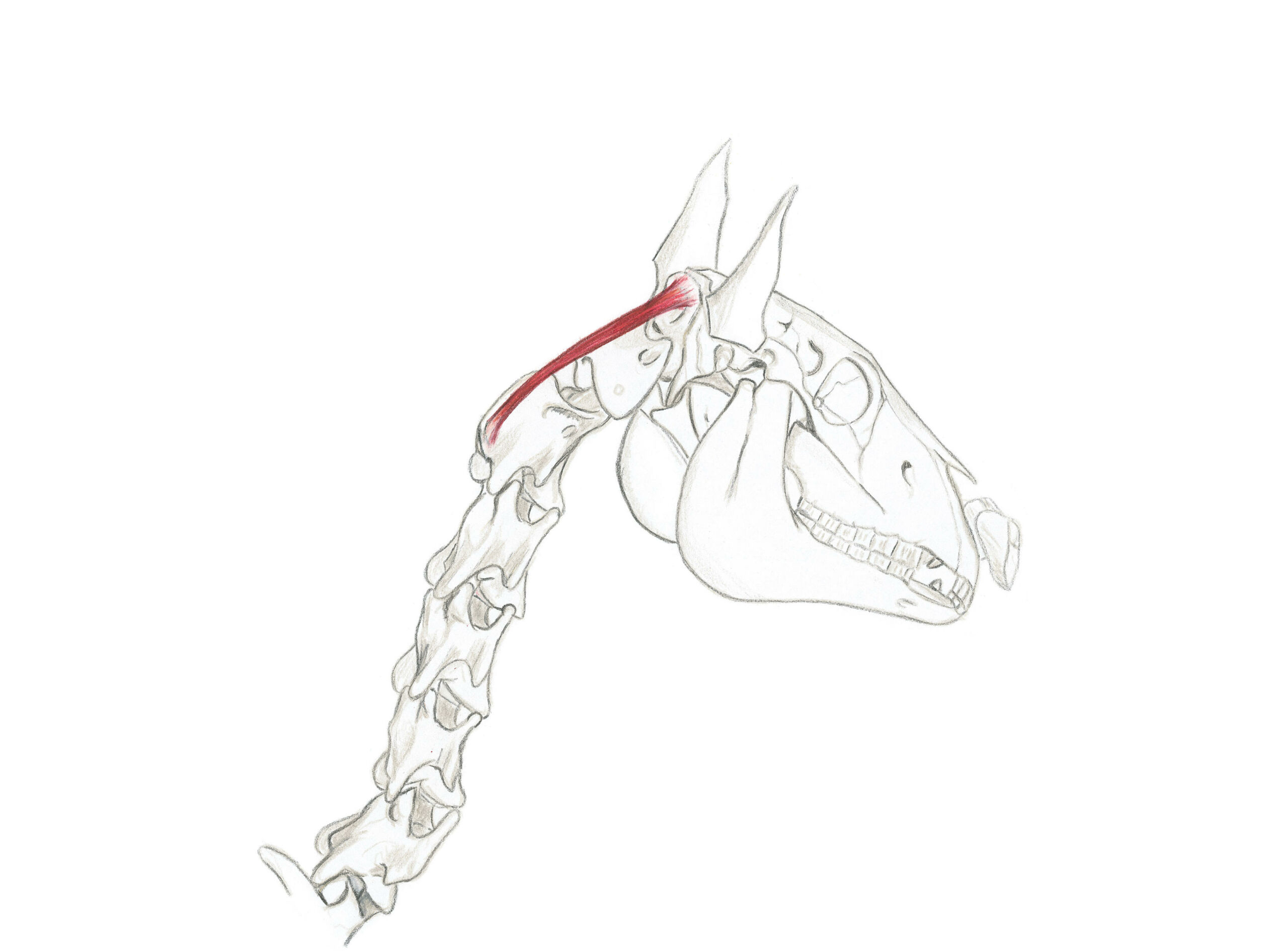
The large dorsal rectus muscle of the head: it is part of the juxta-vertebral muscles of the nape of the neck, the various muscular systems change in the atloid region to act especially on the head, the large dorsal rectus participates in the extension and the inclination of the head.
The rectus abdominis muscle of the head
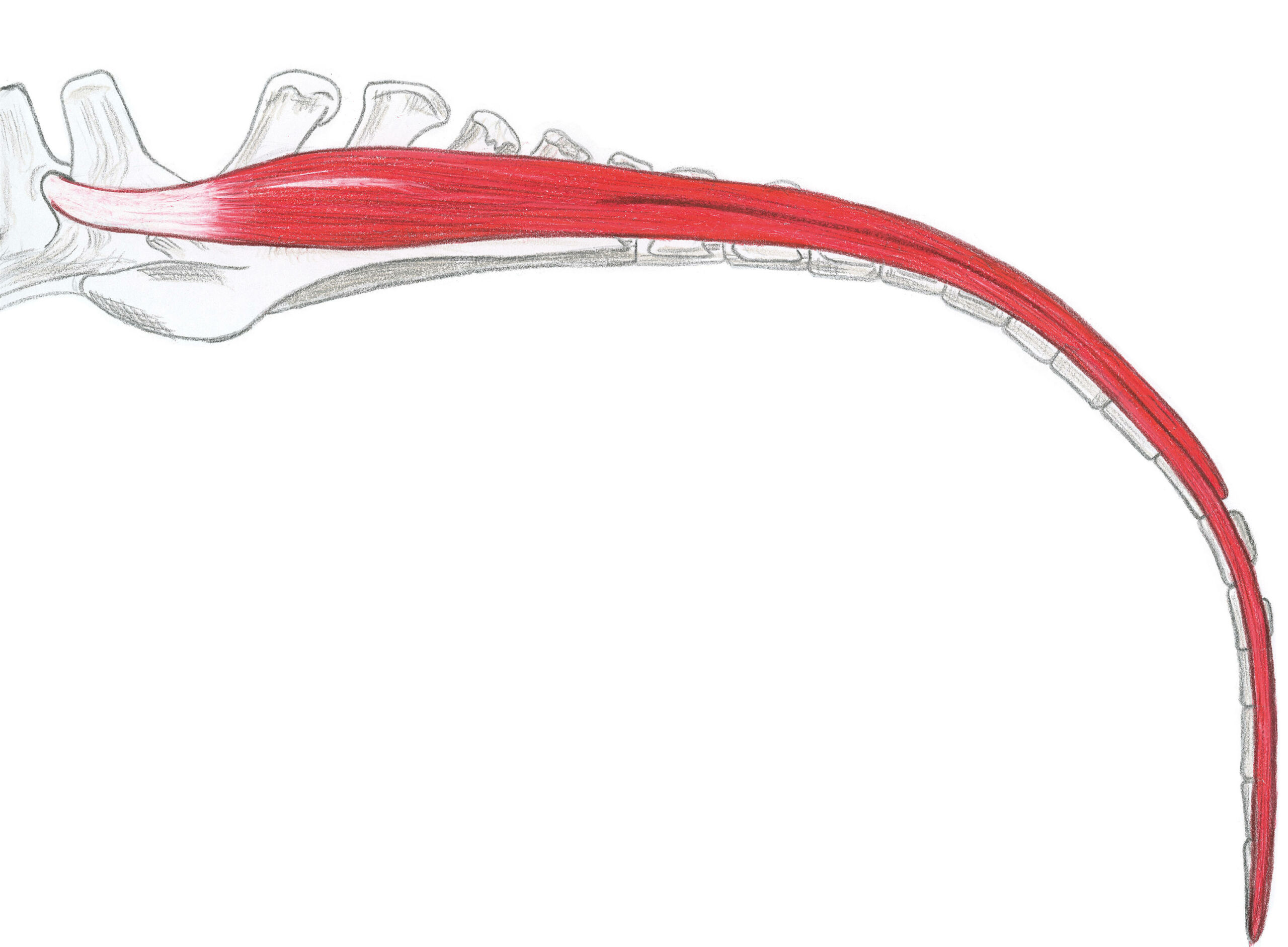
The lateral dorsal sacrococcygeal muscle
Part of the intrinsic tail muscles along with the sacrococcygeals: dorsal-medial, dorsal-lateral, ventral-medial, ventral-lateral and the coccygeal intertransversaries (dorsal and ventral). All the intrinsic muscles are maintained by the coccygeal fascia.
The lateral dorsal sacrococcygeal muscle
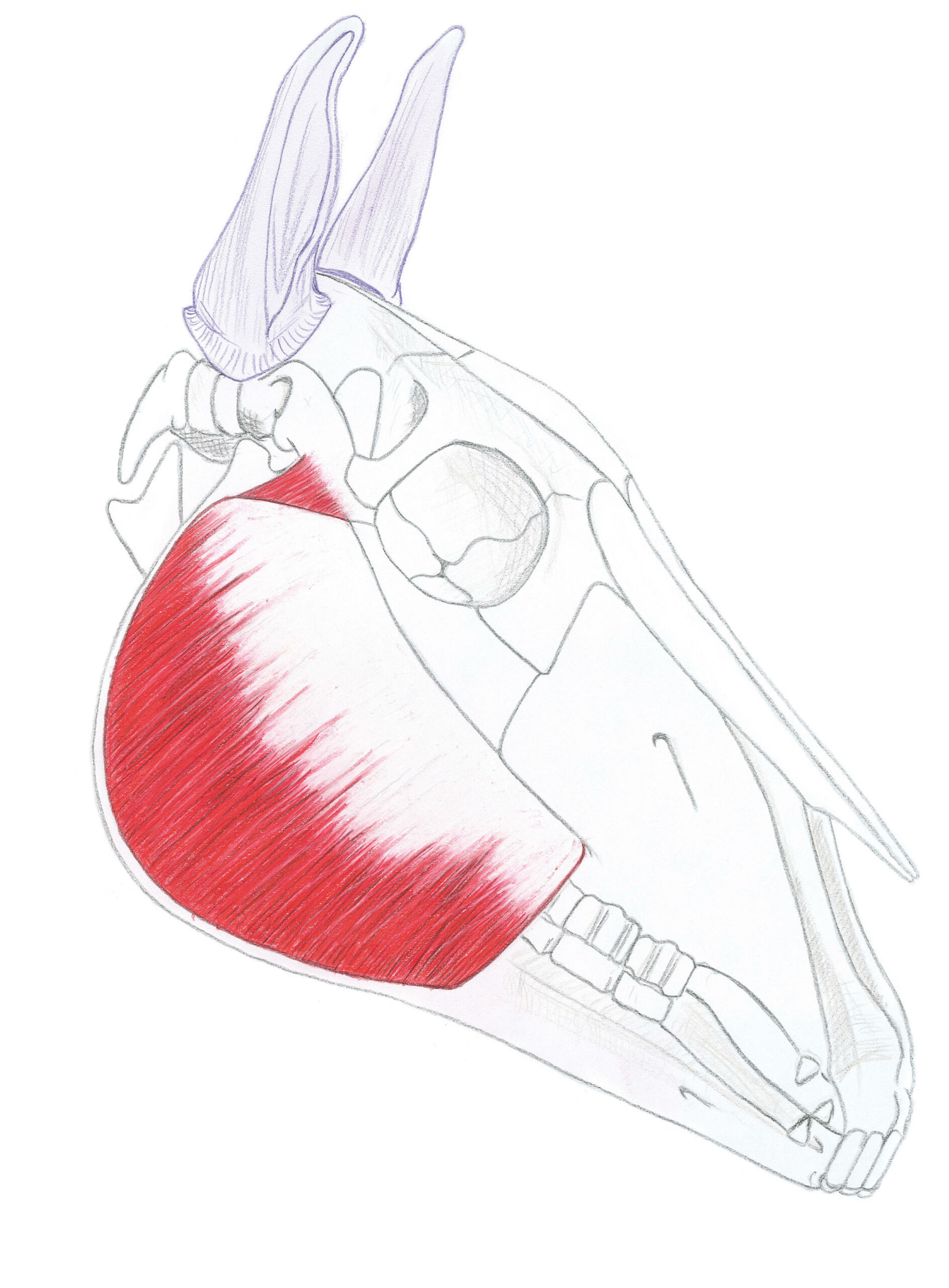
The masseter muscle
The masseter muscle
It gives the cheek area its flat appearance. This region is called the “flat of the cheek” in the horse. It is in two parts; one superficial, the other deep; they are perfectly distinct only in the vicinity of the temporomandibular joint (TMJ).
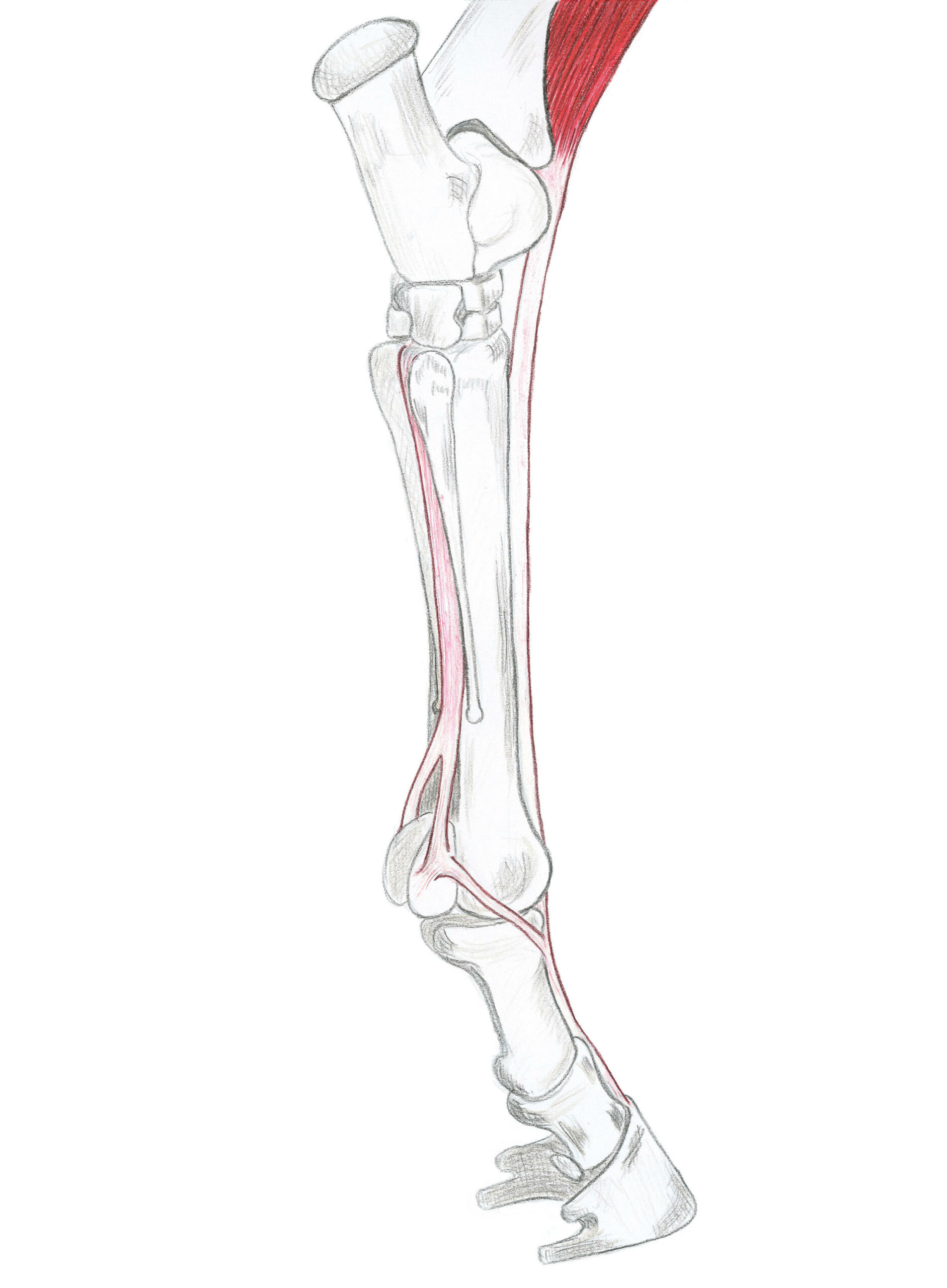
The rope of the hock
It is of complex constitution, particularly powerful in the horse, it takes part in the
tarsal joint angle. It is mainly constituted by the tendon of the sural triceps, in which the participation of the soleus muscle is very weak or even null. It is reinforced by the tendon of the superficial flexor of the finger, which widens opposite the calcaneal tubercle to slide over the end of the tendon of the sural triceps muscle, forming a large fibrous cap; “the calcaneal cap of the plantar perforator”, before continuing on the plantar side of the metatarsus.
The hock cord connects the distal end of the femur and the tuberosity of the calcaneus, a link that maintains the stifle and tarsal joints in constant tension with the 3rd peroneal cord. This connection represents one of the most remarkable adaptations of the limbs to running.